
Diagnostic imaging for dogs and cats
Computed tomography (CT)
CT produces cross-sectional images of the animal’s body and supports detailed assessment of many anatomical regions.
Telephone booking is available daily, 8:00–22:00.
Uses of CT
What can CT assess?
The veterinarian chooses the area to examine according to each patient’s symptoms and needs.
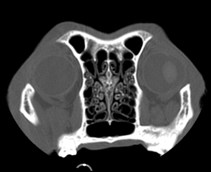
Nasal cavities and sinuses
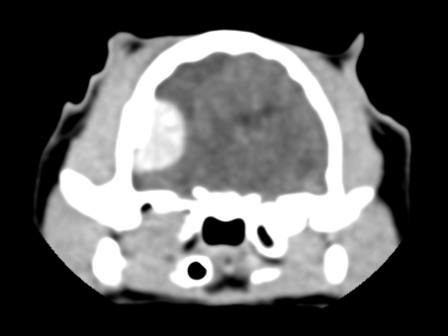
Brain
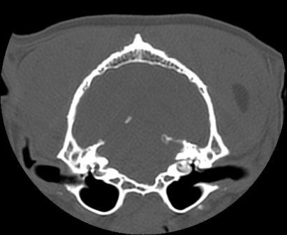
Tympanic bullae
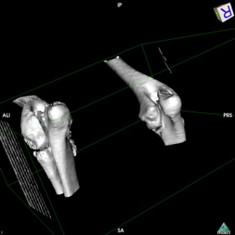
Elbow dysplasia
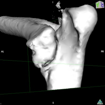
OCD of the joints
Shoulder, elbow, knee and hock joints
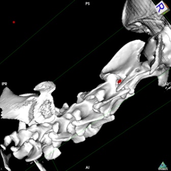
Intervertebral disc disease
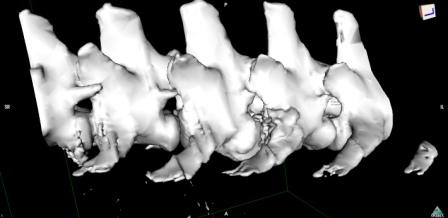
Discospondylitis
Thoracic conditions
CT can help assess structures within the chest.
Before the examination
Confirm preparation with reception
Preparation depends on your pet’s condition and the planned examination. During booking, we will explain which documents and previous test results to bring and any further instructions.
Required information
Radiation safety
Date: 09.10.2025Information on the impact of the clinic’s activities on human health and the environment.
The clinic uses an X-ray unit, among other equipment, for animal diagnostics. The X-ray facility was opened under a permit issued by the President of Poland’s National Atomic Energy Agency.
The clinic monitors exposure of staff and other people through individual and environmental dosimetry carried out by an accredited laboratory. Staff have received radiation protection training from the facility manager.
Appropriate shielding of walls and ceilings, working methods, established processes and procedures, and regular dose readings ensure that the clinic’s activities involving exposure to ionising radiation have no negative impact on human health or the environment.